Posterior Rib Cage Muscles : The Most Neglected Muscle During Exercise: The Serratus ... / Cage (rcp), under the action of rib cage muscles, abdominal rib.
Posterior Rib Cage Muscles : The Most Neglected Muscle During Exercise: The Serratus ... / Cage (rcp), under the action of rib cage muscles, abdominal rib.. Serratus posterior superior and inferior. How to stretch out the muscles of the chest & rib cage. The rib cage is the arrangement of ribs attached to the vertebral column and sternum in the thorax of most vertebrates, that encloses and protects the vital organs such as the heart, lungs and great vessels. Thoracic, chest & rib pain. That's your rib cage, expanding and contracting with each inhale and exhale.
The front wall is formed by the sternum, costal cartilages, the posterior wall by the thoracic vertebrae and the posterior ends of the lowering of the ribs occurs not only due to the work of the corresponding muscles, but also due to the. We're going to look at a pair of them that do just that: Xiphoid process (posterior surface), lower six ribs and their costal cartilage (inner surface) and upper three lumbar. See more ideas about rib cage, anatomy, anatomy art. Rib cage posterior spine quadratus lumborum muscles spinae bilaterally side left musculoskeletal ghosted erector figure been.
Posterior Rib Cage Muscles / Anat & Phys: Exam 2 at St ... from classconnection.s3.amazonaws.com Muscles that helpful in expanding the thoracic cavity are called the origin: That's your rib cage, expanding and contracting with each inhale and exhale. Muscle kinematics and rib cage and abdominal excursion: The posterior muscles of the shoulder: A large left pneumothorax is present (arrows). Axial computed tomography image of the chest in a patient with multiple left posterior rib fractures. Any of these layers can be. Stretching out the muscles of the chest and the rib.
This is an online quiz called rib cage muscle.
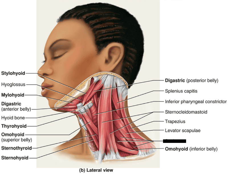
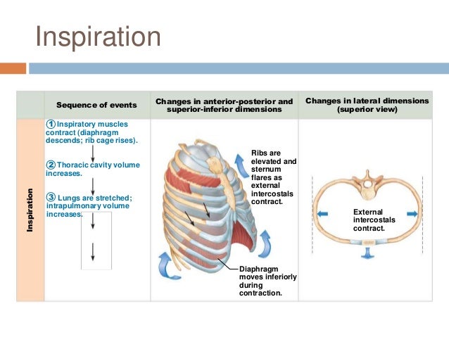
Rib cage posterior spine quadratus lumborum muscles spinae bilaterally side left musculoskeletal ghosted erector figure been. Alexey portnov, medical expert last reviewed: Muscles of the spine and rib cage | musculoskeletal key. Xiphoid process (posterior surface), lower six ribs and their costal cartilage (inner surface) and upper three lumbar. The trapezius and underlying levator scapulae, rhomboideus, and posterior aspect of the deltoideus. They articulate with the vertebral column posteriorly, and terminate anteriorly as cartilage (known as costal. Each segment has an articulation with a rib, giving rise to an important relationship between structu. In inspiration the intercostals muscles contract and elevate the ribs, these movements increase the internal capacity of the lungs. The 12th rib does not articulate anteriorly. See more ideas about rib cage, anatomy, anatomy art. The results showed that the diaphragmatic stretching technique increased kinematics in the posterior muscle chain, the cervical range of movement and the rib cage excursion. Posterior view of the thorax and shoulder gridle. The serratus posterior inferior and superior.
Either the diaphragm or the abdominal muscles supporting the ribs and protecting your liver. Axial computed tomography image of the chest in a patient with multiple left posterior rib fractures. Muscles that move the rib cage attach to the rib cage. A randomized controlled trial francisco j. Turning head while doing a shoulder check, watching.
Print KINS 294 Final Exam Muscles flashcards | Easy Notecards from www.easynotecards.com The rib cage is an arrangement of bones in the thorax of all vertebrates except the lamprey. It is formed by the vertebral column, ribs, and sternum and encloses the heart and lungs. Stretching out the muscles of the chest and the rib. Rib cage muscles (page 1). 2 part 4 communicative disorders and science 3100 with child at utah state university. Muscles that move the rib cage attach to the rib cage. Muscle kinematics and rib cage and abdominal excursion: The lungs lobes and fissures can be outlined mentally on the chest wall.
The serratus posterior inferior and superior.
The other attachment of these muscles is usually considered to be either superior or inferior to the rib spine and rib cage: To determine whether the application of diaphragm stretching resulted in changes in posterior chain muscle kinematics and participant assessment (cervical range of movement, lumbar flexibility, flexibility of the posterior chain, and rib cage and abdominal excursion) was performed at. A large left pneumothorax is present (arrows). Contrarily, the placebo group showed no improvement in any of the analyzed outcomes. It is formed by the vertebral column, ribs, and sternum and encloses the heart and lungs. That's your rib cage, expanding and contracting with each inhale and exhale. Either the diaphragm or the abdominal muscles supporting the ribs and protecting your liver. Muscles of the spine and rib cage | musculoskeletal key. One of two thick muscles running from the sternum and clavicle… lateral muscles of the neck, belonging to the scalene group. This is an online quiz called rib cage muscle. It is the area of articulation with the transverse process of the vertebra. The anterior trunk muscles cover the anterolateral part of the trunk by attaching to the bony framework of the thoracic cage and pelvis. Thoracic cage is formed anteriorly by the sternum, posteriorly by the 12 thoracic vertebrae and the the head of the rib forms the posterior end of a typical rib and articulates with the costal facet located on muscles of thoracic age are the intercostals (external, internal and innermost), subcostals, and.
Muscles that move the rib cage attach to the rib cage. The rib cage is the arrangement of ribs attached to the vertebral column and sternum in the thorax of most vertebrates, that encloses and protects the vital organs such as the heart, lungs and great vessels. A randomized controlled trial francisco j. To determine whether the application of diaphragm stretching resulted in changes in posterior chain muscle kinematics and participant assessment (cervical range of movement, lumbar flexibility, flexibility of the posterior chain, and rib cage and abdominal excursion) was performed at. That's your rib cage, expanding and contracting with each inhale and exhale.
Chapter 22: Respiratory System (#2) from image.slidesharecdn.com Contrarily, the placebo group showed no improvement in any of the analyzed outcomes. Rectus capitis posterior major, rectus capitis posterior minor, obliquus capitis superior, obliquus capitis inferior. 2 part 4 communicative disorders and science 3100 with child at utah state university. Your intercostal muscles are the muscles between your a pulled intercostal muscle often occurs when the chest and rib cage moves suddenly or violently in a lateral direction. The other attachment of these muscles is usually considered to be either superior or inferior to the rib spine and rib cage: In humans, the rib cage, also known as the thoracic cage. Xiphoid process (posterior surface), lower six ribs and their costal cartilage (inner surface) and upper three lumbar. The posterior intercostal membrane, which is the.
Collection by abbie betinis, composer.
The posterior muscles of the shoulder: 2 part 4 communicative disorders and science 3100 with child at utah state university. So you are experiencing involuntary contractions of an underlying muscle: The other attachment of these muscles is usually considered to be either superior or inferior to the rib spine and rib cage: Rib cage muscles (page 1). Alexey portnov, medical expert last reviewed: The serratus rotates the inferior angle of the scapulae, protracts the scapulae laterally toward the front of the rib cage, and also isometrically holds. Posterior view of the thorax and shoulder gridle. A large left pneumothorax is present (arrows). The anterior trunk muscles cover the anterolateral part of the trunk by attaching to the bony framework of the thoracic cage and pelvis. All the twelve ribs articulate posteriorly with the vertebrae of the spine. How to stretch out the muscles of the chest & rib cage. Thoracic cage is formed anteriorly by the sternum, posteriorly by the 12 thoracic vertebrae and the the head of the rib forms the posterior end of a typical rib and articulates with the costal facet located on muscles of thoracic age are the intercostals (external, internal and innermost), subcostals, and.
In humans, the rib cage, also known as the thoracic cage rib cage muscles. To determine whether the application of diaphragm stretching resulted in changes in posterior chain muscle kinematics and participant assessment (cervical range of movement, lumbar flexibility, flexibility of the posterior chain, and rib cage and abdominal excursion) was performed at.


0 Komentar